Boost · UFAT · AugmentDOCX · 48 KB
Chamber Instructions for Use
Rev. 6 · 30 October 2026
Italian original

LIPOSMART ACADEMY
Product documents, scientific learning and training. Find what you need for the system you use.
Choose your system · Find your material · Learn at your pace
DOCUMENT LIBRARY
Rev. 6 · 30 October 2026
Italian original
UPDATED 13 OCTOBER 2026
Video language not confirmed
UPDATED 23 OCTOBER 2026
Video language not confirmed
UPDATED 15 OCTOBER 2026
Video language not confirmed
REV. 9 · 20 NOVEMBER 2025
Italian original
UPDATED 15 OCTOBER 2026
Video language not confirmed
No resources match your selection.
Tell us what you need and which system you use.
Videos open in Google Drive. Contact us if you need access. Use training materials alongside the Instructions for Use.
SCIENTIFIC LEARNING
LEARNING & ILLUSTRATIONS
Choose a topic to browse the illustrations. Open an image for a closer look.
Educational illustrations. Original research microscopy is available in Science & evidence.
Adipose lobules, supporting fibres and small vessels.
Cells, vascular structures and the surrounding matrix.
A closer view of a cell in contact with extracellular matrix fibres.
A conceptual view of communication between neighbouring cells.
A wider view of tissue relationships.
An illustration of signals passing between cells within the matrix.
An illustration of small tissue clusters with their supporting structures.
An illustration of cells separated from their surrounding tissue.
A geometric illustration of how smaller pieces create more surface area at the same total volume.
A simplified cutaway: at rest, filling and elastic return.
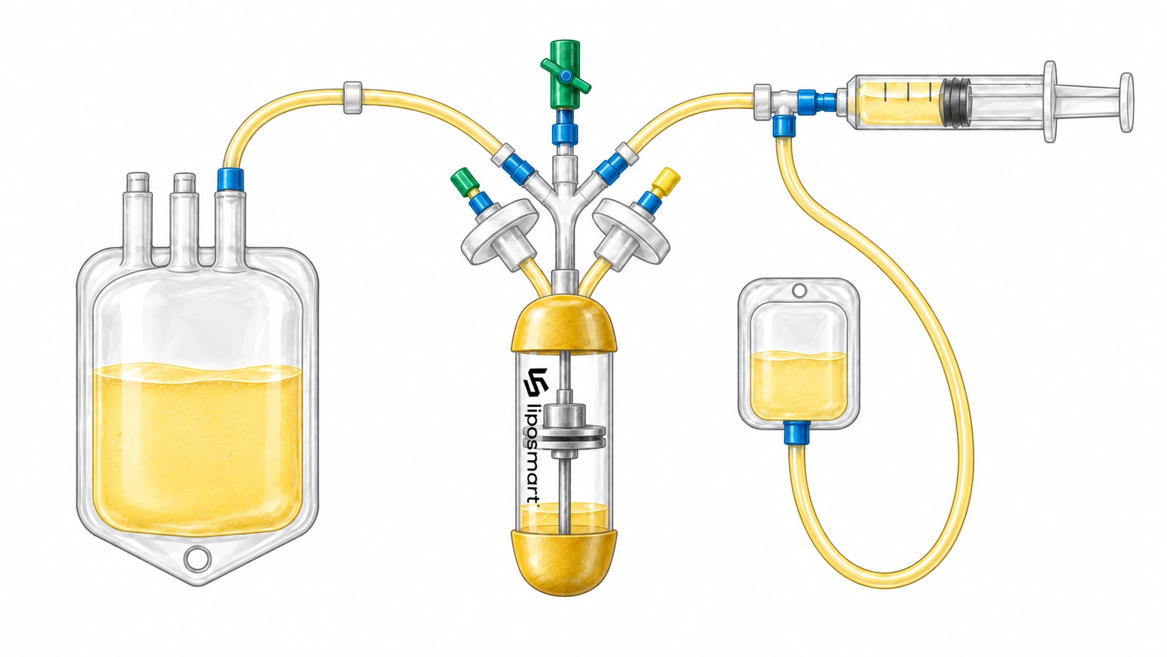
A diagram of the preparation stages, waste collection and tissue recovery.
LIPOSMART
Tell us your country, system and training needs.