Surviving region
Some adipocytes close to the graft edge remained viable. Proximity to the surrounding tissue is central to this model.

SCIENCE / THE THREE-ZONE MODEL
Tissue size is part of the story.
Its living architecture and surroundings matter too.
The three-zone model of fat-graft survival and remodelling adds a biological dimension to the UFAT concept: what happens after tissue is transferred, and why different regions can follow different paths.
Explore the three tissue fates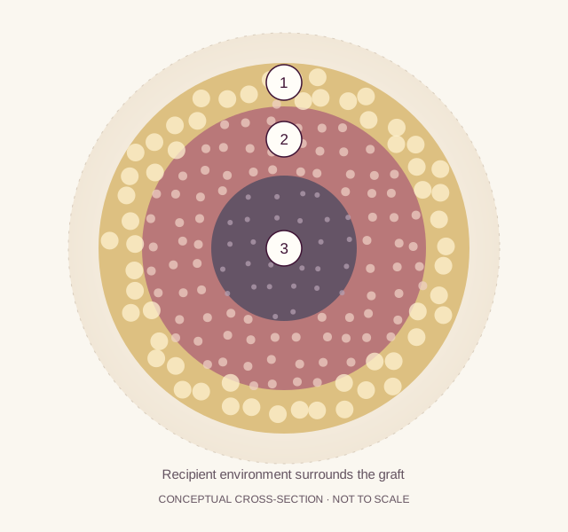
Background science from experimental nonvascularized fat grafts. This diagram is not a liposmart sample or a prediction for every fragment.
ONE GRAFT / DIFFERENT CELL FATES
Eto and colleagues described three spatial regions in mouse fat grafts. Their laboratory studies also found that stromal cells tolerated ischaemic conditions longer than mature adipocytes. [1]
Some adipocytes close to the graft edge remained viable. Proximity to the surrounding tissue is central to this model.
Mature adipocytes were lost, while surviving stromal/progenitor cells contributed to replacement adipocytes. Regeneration here means rebuilding adipose tissue.
In the central region, both cell populations were lost. Subsequent tissue changes could include resorption, fibrosis or oil-cyst formation. [2]
THE RECIPIENT ENVIRONMENT
Before a graft develops a new blood supply, oxygen must reach it by diffusion. Khouri and colleagues modelled how deposit geometry, recipient perfusion and tissue pressure influence oxygen delivery. Their calculations provide a framework, not a universal clinical size threshold. [3]
A prepared fragment and the deposit formed after delivery are different geometries. Packing and coalescence can change the distance oxygen must travel. Recipient vascularity also matters; processing alone cannot determine the environment a graft encounters.
Kato’s mouse study followed remodelling for three months. New adipocytes appeared in the regenerating region, while areas that failed to regenerate followed a different course. These experimental time points are not a human treatment timetable. [2]
READING THE MICRONS
The often-cited 600–1,200 µm range is described as regenerating-zone thickness in a later review. It is not a demonstrated optimal liposmart fragment diameter. [4]
The opening in a device component.
The measured distribution of tissue after processing.
The shape and arrangement of tissue after delivery.
A spatial description of tissue response within a graft.
Eto reported that only some adipocytes within 300 µm of the graft edge survived in the mouse model. This observation is not a minimum fragment-size requirement. [1]
Shih and colleagues describe the regenerating region as 600–1,200 µm thick, citing earlier literature. Reviews differ in describing thickness versus depth from the surface. The original measurement basis and coordinate convention must be resolved before using this range as a quantitative design rule. [4]
For a sphere, diameter is twice its radius. Neither dimension is interchangeable with a layer thickness, and actual adipose preparations are irregular. A filter specification alone cannot establish a recovered particle-size distribution.
Consider the tissue together with the conditions it will encounter.
The UFAT narrative brings cells, microvessels and matrix into the same frame. The three-zone model adds a complementary question: how does the prepared tissue interact with its recipient environment?
For liposmart, this is a scientific rationale for evaluating tissue dimensions alongside viability and retained structure. It does not, by itself, demonstrate preserved function, superior graft retention or a clinical advantage.
Microscopy can show architecture. Demonstrating living cells, functional perfusion and patient benefit requires additional, distinct evidence.
FROM BIOLOGY TO CLINICAL EVIDENCE
This research concerns nonvascularized fat grafts. It does not establish cartilage regeneration, wound healing or outcomes after intra-articular MFAT injection. Those questions need their own indication-specific clinical studies.
Explore the clinical evidence by fieldFOLLOW THE EVIDENCE
Human laboratory material and mouse graft experiments. Three-zone observations; primary abstract reviewed.
Mouse study of remodelling over three months; primary abstract reviewed.
Original mathematical modelling of oxygen delivery and recipient conditions; full text reviewed.
Secondary review, included to trace the 600–1,200 µm terminology. It does not establish a device-specific optimal size.
Scientific content reviewed 12 November 2026. General literature and liposmart-specific reports are identified separately. Original diagram; conceptual and not to scale.